Updated and contextualized version of an article originally published on June 16, 2021
The article retains its original focus by presenting it through a scholarly and accessible perspective, supported by verifiable references.
Authors
- Dr. A. Conte – Biologist
- Roberto Panzironi –Independent researcher
Note editoriali
- First publication: June 16, 2021
- Last update: April 18, 2026
- Version: 2026 narrative revision
Introductory Note
Article previously published and updated following scientific verification and popularization criteria. The text summarizes experimental research and scientific reviews for the general public; it does not replace medical advice or provide personalized clinical recommendations.
IN BRIEF
- Research published in Cell Metabolism showed that, in cellular models and mice, the accumulation and peroxidation of certain PUFAs (including DHA) in acidic tumor microenvironments can induce ferroptosis and slow tumor growth [1].
- Ferroptosis is a form of cell death dependent on iron and lipid oxidation: it is a biological mechanism distinct from apoptosis or necrosis and has therapeutic implications currently under study [2].
- Promising laboratory results do not equate to proof that omega-3 consumption cures or prevents specific cancers in humans; clinical translation requires controlled studies and risk/benefit assessment [5][6].
- Experimental evidence suggests potential combined strategies (PUFA-rich diet + inhibitors of lipid storage or ferroptosis inducers), but these are still preclinical pathways [1][3].
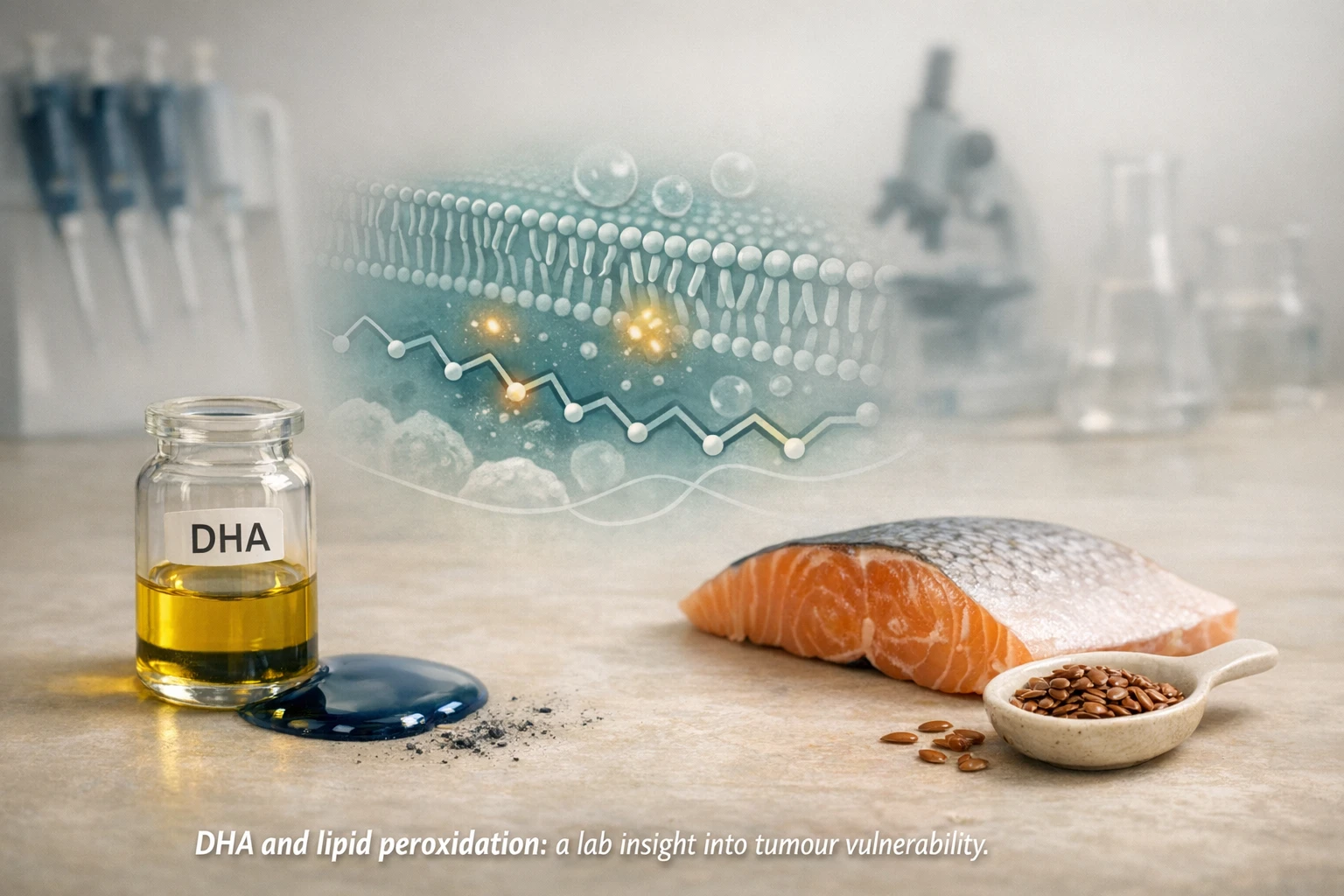
Abstract: what does science say?
The central point emerging from recent literature is that certain polyunsaturated fatty acids (PUFAs) — including DHA, an omega-3 found primarily in fish — can make some cancer cells susceptible to a particular type of cell death called ferroptosis. In experimental models, cancer cells adapted to acidic microenvironments accumulate PUFAs in lipid droplets; once storage capacity is exceeded, these lipids peroxidize, causing membrane damage and cell death. In some mouse studies, PUFA-rich diets slowed tumor growth, and the effect was amplified by drugs that prevent lipid storage in droplets. However, these observations remain preclinical: the response depends on tissue type, the metabolic state of the cells, the iron-antioxidant balance, and other factors of the tumor microenvironment. Therefore, caution is needed before drawing solid clinical or nutritional implications.
The scientific framework: proposed mechanisms
What is ferroptosis?
Ferroptosis is a regulated form of cell death characterized by the lethal accumulation of lipid peroxides: it requires iron and is distinguished from apoptosis and necrosis by morphological and biochemical aspects. This process was first described as a distinct biological entity and has since been linked to metabolic vulnerabilities of cancer cells and other tissues. Sensitivity to ferroptosis depends on three main factors: availability of PUFAs in membranes, presence of catalytic iron, and impairment of antioxidant systems (e.g., the glutathione-GPX4 pathway). [2]
The role of PUFAs and DHA
PUFAs — particularly long-chain polyunsaturated acids like DHA (n-3) and some n-6 — are molecules particularly susceptible to oxidation due to their double bonds. When they accumulate in membranes or in certain lipid pools, their peroxidation can trigger reactive chains that lead to loss of membrane integrity and ferroptosis. Recent experimental studies show that in acidic tumor microenvironments (acidosis), lipid uptake is increased and the ultimate fate of these PUFAs (storage vs. incorporation into membrane phospholipids) affects sensitivity to peroxidation. [1][3]
Experimental evidence and models
Cellular and mechanistic results
A study published in Cell Metabolism used three-dimensional models (spheroids) and cell cultures in acidic conditions to show that exposure to LC-PUFA (long-chain PUFA), including DHA, leads to an increase in lipid peroxidation and spheroid collapse phenomena in relatively short times. The effect intensifies if the formation of lipid droplets (via DGAT inhibitors), which normally serve to buffer peroxidation by storing triglycerides, is blocked. In animal experiments, a diet rich in n-3 LC-PUFA slowed tumor growth compared to a diet rich in MUFA, and the effects were enhanced by co-treatments that promote ferroptosis. These data provide mechanistic consistency between lipid accumulation, peroxidation, and cell damage in experimental tumor contexts. [1]
Results in organisms and limitations
Experiments in model organisms (mice) have shown that diets enriched in LC-PUFA can delay the growth of some tumors, but the effect varies between models and tumor types. Some studies suggest that, in certain contexts, monounsaturated fatty acids like oleate may have a protective effect against ferroptosis, by modifying the lipid composition of membranes and reducing the integration of PUFAs susceptible to peroxidation. These differences highlight that the response is not universal and depends on specific metabolic and microenvironmental networks. [3][4][8]
Clinical context and translation: what is missing
Direct evidence in patients is scarce: most available studies are preclinical or observational and do not demonstrate that increasing DHA or other PUFA intake cures or prevents cancer in humans. Reviews and context analyses highlight that sensitivity to ferroptosis is modulated by numerous factors (redox status, intracellular iron, expression of enzymes like ACSL4, GPX4, FSP1, and lipoxygenase activity) and that ferroptosis itself can have different effects depending on cell type and microenvironment. Translating a nutritional or pharmacological strategy requires controlled clinical trials and safety evaluations, because lipid peroxidation is a potentially harmful process even for healthy tissues. [5][6]
What it means in practice
For the general public, the practical message is one of cautious interpretation. The discovery that, in experimental models, an excess of certain PUFAs can promote the death of cancer cells is important for research but does not authorize considering omega-3s as a "cure" or as an autonomous therapy. Established nutritional recommendations for good cardiovascular and brain health suggest consuming fish rich in omega-3s and a varied diet; however, any dietary modification aimed at oncological therapeutic purposes must be discussed with a doctor. Clinical contexts that evaluate the use of supplements or specific diets as adjuvants to anti-cancer therapies require controlled protocols, monitoring, and an individual risk assessment. [5][6]
Key takeaways
- Ferroptosis is a regulated form of cell death mediated by PUFA peroxidation; it is the subject of intense research in oncology [2].
- Preclinical studies, including the work by UCLouvain, show that DHA and other LC-PUFAs can induce ferroptosis in cancer cells adapted to acidosis and slow tumor growth in animal models [1].
- The effects depend on the metabolic context, the type of PUFA, the presence of defense mechanisms (e.g., lipid droplets, GPX4, FSP1), and the tumor microenvironment [1][5].
- Laboratory results do not automatically translate into clinical recommendations for people: human trials and safety studies are needed [6].
Limitations of the evidence
It is crucial to distinguish between observational evidence, experimental data, and causal evidence in the clinical setting. Many of the cited works are laboratory studies or studies on animal models: they provide information on mechanisms but do not establish that a dietary modification in humans produces the same effect. Observational studies on the association between omega-3 consumption and disease risk can be influenced by confounding factors (lifestyle, other dietary components, socioeconomic status). Furthermore, the biological heterogeneity of tumors means that an intervention that works in one model might not work in another. For these reasons, interpretation requires caution and subsequent clinical research steps after preclinical ones. [2][5]
Editorial conclusion
The discoveries linking the accumulation and peroxidation of PUFAs to ferroptosis in cancer cells open interesting avenues for oncology research, especially as a strategy combined with pharmacological agents that modulate lipid metabolism or the antioxidant capacity of cells. However, it remains crucial not to overinterpret experimental results and to maintain a clear distinction between preclinical evidence and validated therapeutic indications for humans. Future research will need to clarify which tumors, under what microenvironmental conditions, and with what co-treatments can truly benefit from approaches that exploit lipid vulnerability. In the meantime, dietary choices for the population should follow validated nutritional guidelines and consultation with health professionals.
Editorial note
This article has been updated with references to peer-reviewed studies and recent reviews to ensure clarity and accuracy. It is for informational purposes only and does not replace the advice of a doctor or healthcare professional. For therapeutic decisions, always consult a treating physician.
SCIENTIFIC RESEARCH
- Dierge E., Debock E., Guilbaud C., Corbet C., Mignolet E., Mignard L., Bastien E., Dessy C., Larondelle Y., Feron O. Peroxidation of n‑3 and n‑6 polyunsaturated fatty acids in the acidic tumor environment leads to ferroptosis‑mediated anticancer effects. Cell Metabolism. https://doi.org/10.1016/j.cmet.2021.05.016
- Dixon SJ., Lemberg KM., Lamprecht MR., Skouta R., Zaitsev EM., Gleason CE., et al. Ferroptosis: An Iron‑Dependent Form of Nonapoptotic Cell Death. Cell. https://doi.org/10.1016/j.cell.2012.03.042
- Perez M.A., Magtanong L., Dixon S.J., et al. Dietary Lipids Induce Ferroptosis in Caenorhabditis elegans and Human Cancer Cells. Developmental Cell. https://doi.org/10.1016/j.devcel.2020.06.019
- Ubellacker J.M., Tasdogan A., Ramesh V., Shen B., Mitchell E.C., Martin‑Sandoval M.S., et al. Lymph protects metastasizing melanoma cells from ferroptosis. Nature. https://doi.org/10.1038/s41586-020-2623-z
- Lee H., et al. An integrated view of lipid metabolism in ferroptosis revisited via lipidomic analysis. Experimental & Molecular Medicine. https://doi.org/10.1038/s12276-023-01077-y
- Ou Y., et al. The interaction between ferroptosis and lipid metabolism in cancer. Signal Transduction and Targeted Therapy. https://doi.org/10.1038/s41392-020-00216-5
- Environmental Determinants of Ferroptosis in Cancer. Cancers. https://doi.org/10.3390/cancers15153861
- Polyunsaturated fatty acids‑induced ferroptosis suppresses pancreatic cancer growth. Scientific Reports. https://doi.org/10.1038/s41598-024-55050-4